Plant cell partitions outline cell form throughout improvement and are composed of interlaced carbohydrate and protein networks. To get the very best reside photographs nevertheless there are a number of elements to contemplate when selecting a mobile stain label or dye.
 Mcf 7 Most cancers Cell Staining Cell Nuclei Stained With Dapi Blue 1 Obtain Scientific Diagram
Mechanisms of antigen.
Cell imaging dyes labels stains. Differentiation and development management. It’s extensively used to establish proliferating cells and labels cell strains and first cell cultures in vitro and likewise cells in vivo. Though not as vibrant because the important Hoechst stains for DNA DAPI has better photostability.
Abberior LIVE dyes are designed for STED and confocal microscopy in residing cells. Tubulin Tracker dyes for live-cell microfilament staining. Howdy Bio presents prime quality but reasonably priced reagents for tissue and cell imaging for clear reproducible staining leads to your immunohistochemistry IHC and stuck and reside cell imaging experiments.
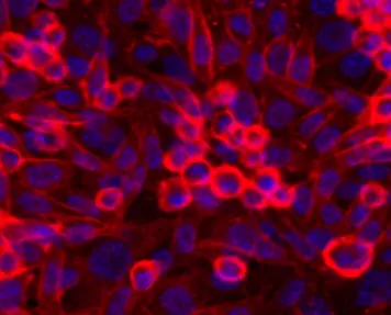
Nuclear staining of fastened and permeabilized U2OS cells utilizing NucBlue Fastened reagent a type of DAPI. Fluorescence labeling has revolutionized live-cell imaging. Many fluorescent dyes are commercially out there and stain a wide range of residing cell organelles such because the nucleus 11 12 mitochondria 13 lysosomes 14 and endoplasmic reticulum 15.
Labels stains dyes BrdU 5-Bromo-2-deoxyuridine is a thymidine analog which is integrated into DNA throughout DNA replication throughout S-phase of cell cycle. Taxolpaclitaxel and taxoteredocetaxel conjugates are used for endpoint assays of cytoskeletal conduct in reside cells to offer intense staining of polymerized tubulin. These particular stains are appropriate counterstains to antibodies to assist the identification of location-specific targets of curiosity inside the cell.
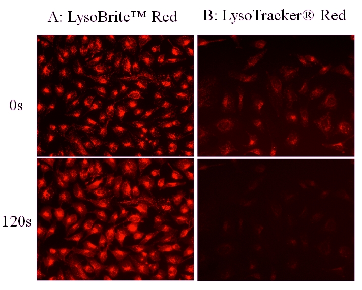
Decrease background than LyosoTracker dyes. These distinctive dyes mix highest brightness and photostability with easy live-cell labeling. Organelle visualization with organelle-selective stains or dyes is a key instrument in fluorescence imaging of cells and tissues.
Fluoresces inexperienced for eukaryotic DNA purple for RNA and orange for prokaryotic cells. 7-AAD – membrane-impermeable purple fluorescent dye that binds DNA. In distinction RNA-specific dyes for staining reside cells are hardly ever out there.
LipidSpot stains present minimal background staining of mobile membranes or different organelles in contrast to conventional dyes like Nile Crimson. For instance inexperienced fluorescent protein GFP and its derivatives. Browse successfully by means of our intensive catalog by choosing applicable filters on the left for attributes resembling product.
Additionally stains acidic organelles resembling lysosomes. 18 rows Reside-or-Dye NucFix Crimson is a novel cell membrane impermeable dye that particularly stains. LipidSpot 488 has excitation round 430 nm and might be excited equally effectively at 405 nm or 488 nm.
As a result of lipophilic cell monitoring dyes resembling PKH26 PKH67 and CellVue Claret can be utilized to label nearly any cell they’ve enabled most cancers biologists to trace all kinds of tumor and immune cell features in vitro and in vivoThese embrace. Fluorescent dyes have lengthy been used to label plant cell partitions enabling optical microscopy-based interrogation of cell wall construction and composition. Some generally used cell staining dyes.
LysoView dyes label lysosomes in reside cells and can be found with blue inexperienced seen purple and far-red fluorescence. These stains embrace Hoechst and 46-diamidino-2-phenylindole DAPI that are used on this article. Researchers can save as much as 50 on cell imaging reagents dyes labels stains from Howdy Bio – they’re round half the worth of different suppliers.
These stains embrace Hoechst and 46-diamidino-2. Nonetheless the precise cell w. We provide ready-to-use probes for DNA tubulin and lysosome labeling.
Non-toxic for real-time live-cell imaging. Reside cell imaging of mobile mitochondria. In cells it stains lipid droplets with vibrant inexperienced fluorescence detectable within the FITC channel.
No-wash fluorescent staining of lysosomes in reside cells. Labels Dyes Stains A large assortment of chemical compounds and reagents can be found to be used in biochemistry molecular biology and cell biology. Reactive Dyes and Stains Illuminating your Outcomes A particular mixture of reagents comprised of remarkable fluorescent molecular probes together with CELLESTIAL dyes for.
I wish to stain two several types of cell strains with totally different coloured dye and wish to examine their interplay in a selected surroundings. CellVue fluorescent imaging kits use proprietary labeling know-how to stably incorporate fluorescent dyes containing lengthy aliphatic hydrocarbon tails into lipid membranes 1They are helpful for researchers working in all points of science and know-how the place fluorescently-labeled cells andor tissues are required. The 2 lengthy 18-carbon chains insert into the cell membrane leading to particular and steady cell staining with negligible dye switch between cellsXenoLight DiR together with PerkinElmers IVIS imaging methods can be utilized for non.
XenoLight DiR – Monitor Cells In Vivo Non-Invasively. Fluorescently Label Cells or Tissues for in vivo or in vitro Research. These rely on the imaging method getting used in addition to organic query being requested.
Lifeless cells purple are labeled with a cell-impermeant dye DeadRed reagent and reside cells inexperienced are stained with calcein. I’m going to make use of a reside cell imaging platform and it. A number of fluorescent stains can be found that label DNA and permit straightforward visualization of the nucleus in interphase cells and chromosomes in mitotic cells.
Imaging dyes stains labels. A number of fluorescent stains can be found that label DNA and permit straightforward visualization of the nucleus in interphase cells and chromosomes in mitotic cells. Acridine Orange – cell-permeable nucleic acid dye.
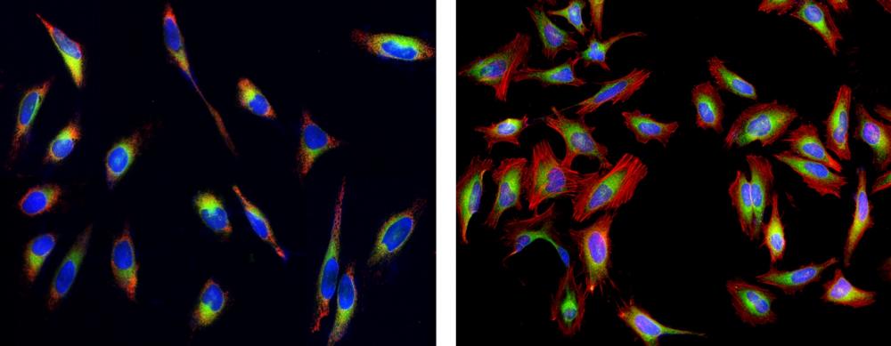
XenoLight DiR is a lipophilic close to infrared fluorescent cyanine dye excellent for staining the cytoplasmic membrane. A Reside HeLa cells co-stained with. Labeling the Nucleus with Fluorescent Dyes for Imaging.
Proliferation of stem and progenitor cells. No transfection and no washing steps are wanted.
![]() Cell Monitoring Crimson Dye Equipment Longer Cell Staining Dmso Free Ab269446
Cell Monitoring Crimson Dye Equipment Longer Cell Staining Dmso Free Ab269446
 Reside Or Lifeless Fixable Lifeless Cell Staining Equipment Orange Fluorescence Aat Bioquest
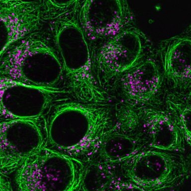
Viafluor Reside Cell Microtubule Stains Biotium
 Cell Navigator Reside Cell Endoplasmic Reticulum Er Staining Equipment Inexperienced Fluorescence Aat Bioquest
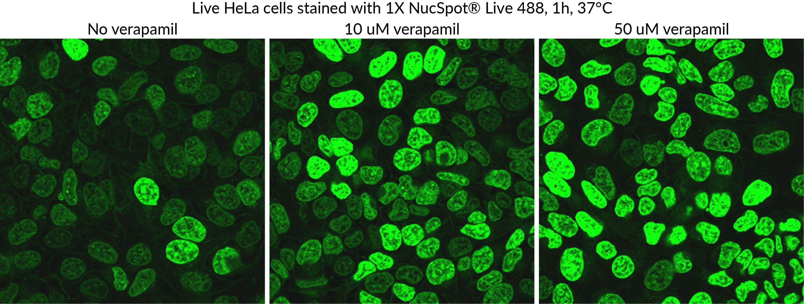
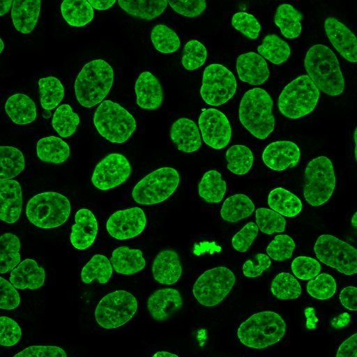 Nucspot Reside Cell Nuclear Stains Biotium
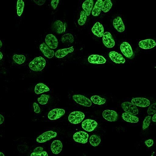 Nucspot Reside Cell Nuclear Stains Biotium
 Cell Viability As Decided By Fluorescein Diacetate Propidium Iodide Obtain Scientific Diagram
 Cell Imaging Dyes Labels And Stains
 Nbd Ldl cholesterol Staining Dye Equipment Ethanol Free Ab269448 Abcam
 Qdot Label Conjugates For Cell Tissue Staining Thermo Fisher Scientific Cell Fluorescence Microscopy Thermo Fisher
 Nuclear Staining For Reside Cell Imaging And Fastened Cells Biocompare Com Equipment Reagent Evaluate
 Cell Navigator Reside Cell Endoplasmic Reticulum Er Staining Equipment Crimson Fluorescence Aat Bioquest
 Nucspot Reside Cell Nuclear Stains Biotium
![]() Cell Monitoring Crimson Dye Equipment Longer Cell Staining Dmso Free Ab269446
Cell Monitoring Crimson Dye Equipment Longer Cell Staining Dmso Free Ab269446
 Cell Navigator Lysosome Staining Equipment Crimson Fluorescence Aat Bioquest
 Bis Azepanyl Rhodamines Potomac Dyes As Inside Membrane Stains Obtain Scientific Diagram
 Cell Navigator Mitochondrion Staining Equipment Deep Crimson Fluorescence Aat Bioquest
 Cell Staining Dyes Biocompare Com
 Reside Cell Fluorescent Organelle Dyes And Stains